Chronic kidney disease led to the destruction of phalanges in a Thai patient.
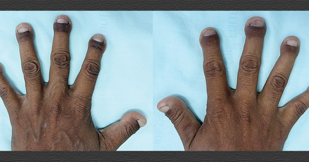
- The patient with chronic kidney disease had destroyed distal phalanges of the fingers.
- The X-ray revealed almost complete disappearance of the phalangeal nodules and osteodystrophy.
- The condition is associated with a disturbance in mineral metabolism due to prolonged nephropathy.
- The authors call for regular monitoring of biochemical indicators and therapy.
A 46-year-old man was admitted to one of the hospitals in Thailand, who had experienced gradual painless shortening and rounding of his fingertips over the past few years. According to doctors, the changes began about ten years ago when the patient was diagnosed with chronic kidney disease caused by obstructive nephropathy and the presence of urinary stones.
The patient's kidney disease developed irregularly: he visited doctors only episodically, which led to the accumulation of metabolic disorders characteristic of prolonged renal failure. Such disorders often cause disturbances in mineral metabolism, which in turn leads to the development of kidney-related bone diseases.
During the examination, doctors noted short, broad, and club-shaped fingertips, while similar but less pronounced changes were observed on the toes. X-ray examination of the hands showed resorption of the tuberosities of all distal phalanges, leaving only small proximal remnants. Additionally, the images revealed osteopenia, vascular calcification, chondrocalcinosis, and osteolytic lesions at the distal ends of several metacarpal bones.
These changes correspond to the characteristic signs of fibrous osteodystrophy – a form of kidney-related bone disease, where the balance of calcium and phosphorus is disrupted, leading to increased resorption of bone tissue and its replacement with fibrous tissue. The condition is often accompanied by bone pain, increased fragility of the skeleton, and, as in this case, deformity of the limbs.
The case was described by doctors Navarat Pengpong from Phrabokklao Hospital and Thiti Snabbun from Chulalongkorn University in The New England Journal of Medicine. The authors noted that such extreme forms of renal osteodystrophy are rare but can occur with prolonged lack of adequate control over phosphate levels, parenteral vitamin D preparations, and other aspects of chronic kidney disease treatment.
Experts emphasize the importance of regular monitoring of biochemical indicators in patients with chronic nephropathy, as well as timely adjustments to diet, phosphate binders, and active forms of vitamin D. Without such therapy, the risk of developing severe bone complications, including phalanx destruction, significantly increases.
The case demonstrates how the systemic consequences of kidney failure can affect even the most remote parts of the body. It serves as a reminder of the need for a comprehensive approach to the treatment of chronic kidney disease, including both nephrological and orthopedic care.
Source: N+1